
Advanced light microscopy is primarily served by a LEICA SP8 Upright CLS/multiphoton/FLIM microscope, and a Keyence-VHX7000 digital microscope., and a Zeiss Axiovert reflected light microscope/metallograp. There are also a range of other smaller LMs, including a Leica epifluorescent stereo microscope (FM/LM) for higher level routine stereo imaging and many small desktop microscopes for casual use.
Email Dr. Tingting Gu for advanced light microscopy consultations or for materials microscopy and metallography contact Dr. Julian Sabisch.
Log into iLab to see the schedule and availability.
For a graphical overview of our ALM capabilities, refer to the SRNML ALM Capabilities (PDF).
We're happy to answer any questions and work with you to acquire new capabilities.


Located in Richards Hall room 9 | Check Leica SP8 confocal Availability
The flagship optical microscope at OU; this microscope is a dual system with both visible laser scanning confocal microscope and multi-photon on a single platform. It is an upright system with motorized stage that is ideal for traditional slide mounts and small animal imaging.
Visible laser:
GaN laser: 405nm
Argon laser: 458, 476, 488, 496
DPSS laser: 561nm
HeNe: 633nm
Multi-photon laser:
Coherent Chameleon Ti: Sapphire laser: tunable from 680nm to 1080nm
Repetitive rate: 80MHz
3 PMT detectors: robust photomultiplier tube (PMT) detectors for bright signals.
3 HyD detectors: highly sensitive hybrid GAsP/APD (HyD) detectors for low light imaging.
Dry objectives
5X 0.15 NA Air WD 13.70 mm HCX PL Fluotar
10X 0.30 NA Air WD 11.00 mm HCX PL Fluotar
Dipping objectives
25X 0.95 NA water WD 2.5mm HCX IRAPO L
40X 0.80 NA water U-V-I WD 3.30 mm HCX APO L
63X 0.90 NA water U-V-I WD 2.2mm HCX APO L
Immersion objectives
20X 0.75 NA Oil, Glycerol WD 0.68 mm HCX PL APO CS2
40X 1.10 NA Water WD 0.65 mm HCX PL APO CORR CS2
63X 1.30 NA Glycerol WD 0.22 mm HC PL APO CORR CS2
63X 1.40 NA Oil WD 0.14mm HC PL APO
100X 1.40 – 0.70 NA Oil WD 0.09mm HCX PL APO CS
DAPI, FITC, TRITC
FiltersBeam splitter Emission CH1 Emission CH2
FITC/TRITC 560 525/50 585/40
CFP/YFP 505 483/32 535/30
PicoQuant TSCPC system 2 channels
Leica Application Suite-Advance Fluorescence (LAS AF)

Contact: Dr. Tingting Gu
The Keyence digital microscope is both easy to use and powerful. The microscope features transmitted and reflected light sources, a fully motorized stage, software for 2D / 3D measurements, montaging, Z-stacking, and many other acquisition/ analysis options. We also have polarizer and analyzer options for reflected and transmission polarized light microscopy.


The Zeiss Axiovert 200M is available free of charge to OU researchers
Location: NML 209 | Check Zeiss Axiovert 200M Availability
Low skill curve for use, with a wide range of features and imaging modes:

Location: NML 207 | Leica MZ 10F Availability
Stereomicroscope with switchable fluorescent or bulb sources and digital imaging.
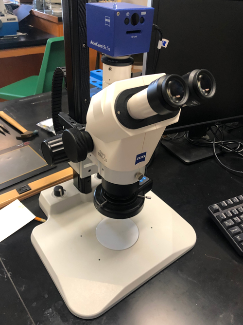
Location: NML 206 | Zeiss 2000-CS Availability
Very easy-to-use stereomicroscope with ring light and color digital imaging system with calibrated scale, 6.5x - 62.5x magnification.
Available to use for no charge.
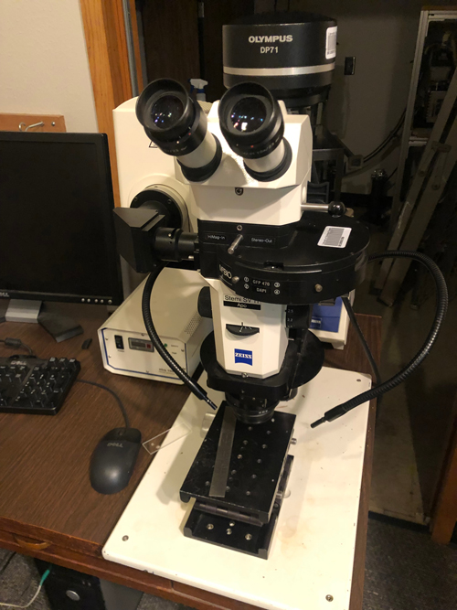
Location: NML 205 | Zeiss Stemi SV11 Availability
Stereomicroscope with switchable fluorescent or bulb sources and basic digital imaging.
Available to use for no charge.
