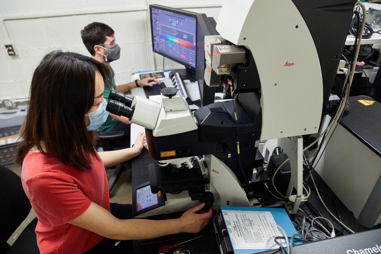
The Samuel Roberts Noble Microscopy Laboratory, the core microscopy facility of the University of Oklahoma-Norman, offers access to instrumentation, training, and service.
Our mission is to provide effective and efficient access to core imaging and analytical characterization technologies. We welcome all OU-Norman, OUHSC, and OMRF student, faculty, and staff researchers at our centralized location the OU-Norman main campus. Clients from other universities, foundations and industry are welcome to use this equipment as well, and such work may be done on an invoice or contract basis.
In addition to instrument access, SRNML personnel offer advice, hands-on training, education, and research collaboration. We have a range of sample preparation equipment available; our staff work with users and provide training. The SRNML aims to promote, enable, and encourage cutting-edge education and research using the core’s instrumentation. Please see our Using SRNML page to get started, and don’t hesitate to contact us.
Become a User, Take a Course, Schedule a Visit or Consultation, and Participate in events
As an OVPRP-supported core facility, SRNML expects acknowledgment (and co-authorship as appropriate) resulting from technical support and collaboration with SRNML personnel.
The following language should be used (or modified as appropriate):
"Data collection was performed at the Samuel Roberts Noble Microscopy Laboratory (SRNML), a University of Oklahoma (OU) core facility supported by the OU Vice President for Research and Partnerships (OVPRP)."
SRNML offers an Acknowledgment Incentive Program
Senior corresponding authors may receive a $100 credit toward future SRNML facility use for published manuscripts with SRNML acknowledgment or co-authorship.
Terms and Conditions of the SRNML Acknowledgment Incentive Program:
We send email updates regarding events, instrument and lab resources, opportunities, etc. from time to time. If you would like to be included in our mailing list, please submit your email address below.
Please join us for an Imaris 3D image analysis workshop hosted by the University of Oklahoma, Samuel Roberts Noble Microscopy Facility. We'll demonstrate how advanced image analysis tools can accelerate your scientific research while delivering more robust, reliable results. The workshop features an Imaris seminar highlighting the latest AI-enhanced analysis capabilities, plus the opportunity to book a 1:1 session with an Imaris specialist to learn how to get the most out of your own microscope images.
We will participate in the annual Kids Night with Microscopes on Tuesday, November 11, from 6–8 PM at The Well in Norman and organized by the Oklahoma Microscopy Society (OMS). OMS invites all science and microscopy enthusiasts to join us for this engaging, hands-on evening of discovery and fun for families, students, and community members of all ages. This popular outreach event regularly draws families and children from across the Oklahoma City metro area, offering an inspiring opportunity for young minds to explore the hidden world of science and technology.
We are thrilled to announce that our newly purchased Zeiss LSM 780 with Airyscan Super-Resolution Module is now fully installed and ready for use! This powerful upgrade expands our imaging capabilities, enabling researchers to achieve higher resolution, improved sensitivity, and faster acquisition for advanced biological and materials science applications. The Airyscan technology delivers super-resolution imaging beyond the diffraction limit while maintaining exceptional signal-to-noise ratios, ideal for live-cell imaging and fine structural analysis.
Our Zeiss Neon 40 EsB dual beam field emission scanning electron microscope (SEM) installed in the Samuel Roberts Noble Microscopy Laboratory (SRNML) recently received an upgraded Energy Dispersive Spectroscopy (EDS) system, allowing greater signal detection, providing improved accuracy, sensitivity, and throughput along with a new Windows 11 PC and an up-to-date software package (Aztec).
SRNML is acquiring the first super-resolution inverted confocal laser scanning microscope for the broader OU-Norman campus community and beyond. The Zeiss LSM780 with Airyscan system will be available through SRNML's user programs and courses, this spring. The acquisition was possible thanks to support from the OVPRP and the Materials Science and Engineering Ph.D. program, funded through an internal proposal led by PIs Stefan Wilhelm, Tingting Gu, and Andy Elwood Madden. The instrument will feature Airyscan superresolution capability, able to reach ~1.7 times improvement (to ~120 nm laterally, ~350 nm axially) over our current light microscopy capabilities. The system includes an incubating enclosure, ideal for imaging of dynamic, live cell processes. As part of the user facility, this instrument will enable state-of-the-art research, education, and training in advanced materials science, and life/health science and engineering. We anticipate the system will be installed and made available for users later this spring, check back or ask with questions or to get updates!
The votes are in, and the Oklahoma Microscopy Society 2024 Ugly Bug winners have been announced! Congratulations to students and teachers from Mazie Elementary, Finley Reese Elementary, Lone Grove Intermediate, Silo Elementary, Casady School, and Washington Elementary! Click for more information.
Join us in Norman for the Oklahoma Microscopy Society spring meeting, 'A year of Firsts for Oklahoma Electron Microscopy'! Dr. Peter Ercius, the Interim Director of the National Center for Electron Microscopy is the keynote speaker, and we’re hoping to do tours of our new Tundra cryo-TEM and JEOL aberration-corrected STEM labs, in addition to committed faculty talks by Oklahoma leaders in these areas: Dr. Rakhi Rajan, Dr. Iman Ghamarian, and Dr. Ritesh Sachan. There will be vendors displaying the latest innovations in microscopy available for discussions. Most importantly, students can submit abstracts to compete for the Timpano award, offering the winner funds to attend the national Microscopy and Microanalysis Conference, a student poster session with cash prizes, and a Best Micrograph Contest, also with cash prizes. See okmicroscopy.org for additional details and to register.
With the award of an NSF MRI and support from the OVPRP, OU has purchased a JEOL NeoARM 200kV aberration corrected STEM materials specific microscope. This state-of-the-art instrument will allow researchers to probe the structures of materials down to an ultimate resolution of 70 pm. With a DECTRIS Ela hybrid-pixel direct electron camera coupled to a CEOS energy filter / EELS, electronic structures of thin films, energy and battery materials, and other semiconductor devices will be able to be investigated on the atomic level. Additionally a high throughput EDS detector allows for quick chemical mapping of all samples imaged on the microscope, leading the way for researchers to quickly investigate all aspects of their materials down to the atomic level. Room B21 Lin Hall has been prepared to house the microscope. Delivery is expected on Tuesday, March 25, and installation will proceed immediately afterwards. We hope to have the instrument ready to support users before the end of the spring semester, stay tuned!
You are invited to attend the research presentations for the Graduate Certificate in Microscopic Imaging & Technology program Thursday Dec. 12th at 10 am - 12 pm at SRNML conference room. This is an opportunity for our students in the certificate program to present their research work related to microscopes or microscopic applications. Your presence will be a great support to our students and our graduate certificate program.
The fall semester is coming to an end, and at SRNML is means that students taking the TEM lab course will be giving their final presentations. The students have been working hard all semester to get TEM data for their research projects after learning how to use our high resolution 2010F microscope. Please join us in the SRNML conference room at 9am on Dec 10th to see all the great work they have been doing!
Prof. Tony Wilson, University of Oxford (Emeritus), Past President of the Royal Microscopy Society and past Editor of the Journal of Microscopy, will be presenting the lecture "Confocal Microscopy Today". All are welcome Monday, December 9th at 1 pm in the SRNML conference room. Prof. Wilson was a key developer of the laser confocal scanning microscope and structured illumination microscopy. We hope to see you there to welcome one of the great pioneers of confocal microscopy!
I am pleased to announce that the ThermoFisher Tundra cryo-TEM, a first in the State of Oklahoma, has been installed, inspected, tested, and is now available for trained users. In celebration of this joint venture and incredible endeavor, we would like to invite you to attend our Grand Opening Event at the Stephenson Life Sciences Research Center on Thursday, December 5, 2024, at 4 pm. The celebration will take place on the 1st Floor North Lounge. We will celebrate with some refreshments, a formal ribbon cutting, a few words to commemorate this event, and small group tours of the facility.
Our workhorse JEOL 2010F with an ultimate resolution of 1Å was recently upgraded with a Nanosprint 12 scientific CMOS camera. This lens-based system allows for high doses and exceptional sensitivity on a wide range of samples. Coupled with the microscope’s STEM and EDS capabilities, new technique development opportunities abound to extend the lifespan of our existing toolset. Reach out to Julian and try it out! Image and corresponding diffraction pattern of gold nanoparticles taken by Julian using the new AMT camera.
This easy-to-use reflected light materials microscope / metallograph is now available for use at no charge to users. Microscope capabilities include: fully motorized xyz stage, 1229 x 968 resolution camera, large area montaging and automated image stitching., brightfield and darkfield imaging needed for grain size analysis and surface quality measurements respectively , up to approximately 3000x fully optical magnification, and Axiovision Pro software for acquisition and analysis.
In addition to the reflected light polarizer, our Keyence digital microscope now has a fully rotatable polarizer/ analyzer setup for transmitted light microscopy. Thanks to several OU faculty, the OU Center for Quantum Research and Technology, OptoKhemia Analytical, and particularly the Furis research group for making this possible.