Skip Navigation

Researchers Publish Findings from ‘Super-Resolution’ Imaging Technology

May 9, 2023

Researchers Publish Findings from ‘Super-Resolution’ Imaging Technology

Stefan Wilhelm, Ph.D., assistant professor in the Stephenson School of Biomedical Engineering

Led by Stefan Wilhelm, Ph.D., assistant professor in the Stephenson School of Biomedical Engineering at the University of Oklahoma, a team of researchers from the Gallogly College of Engineering at OU, OU Health Sciences Center and Yale University recently published an article in ACS Nano that describes their development of a super-resolution imaging platform technology to improve understanding of how nanoparticles interact within cells.

As technology-driven capabilities in engineering and healthcare are ever increasing, scientists and engineers are developing new technologies to advance the future of health. One such area, nanomedicine, explores the use of nanoparticles for drug delivery in the body to fight against infectious diseases or cancer. The assessment of these nanomedicines in cells, tissues and organs is often performed by optical imaging, which can have a limited quality of imaging resolution. New imaging technologies are needed to see nanoparticles in their 3-D ultrastructural context within biological tissues.

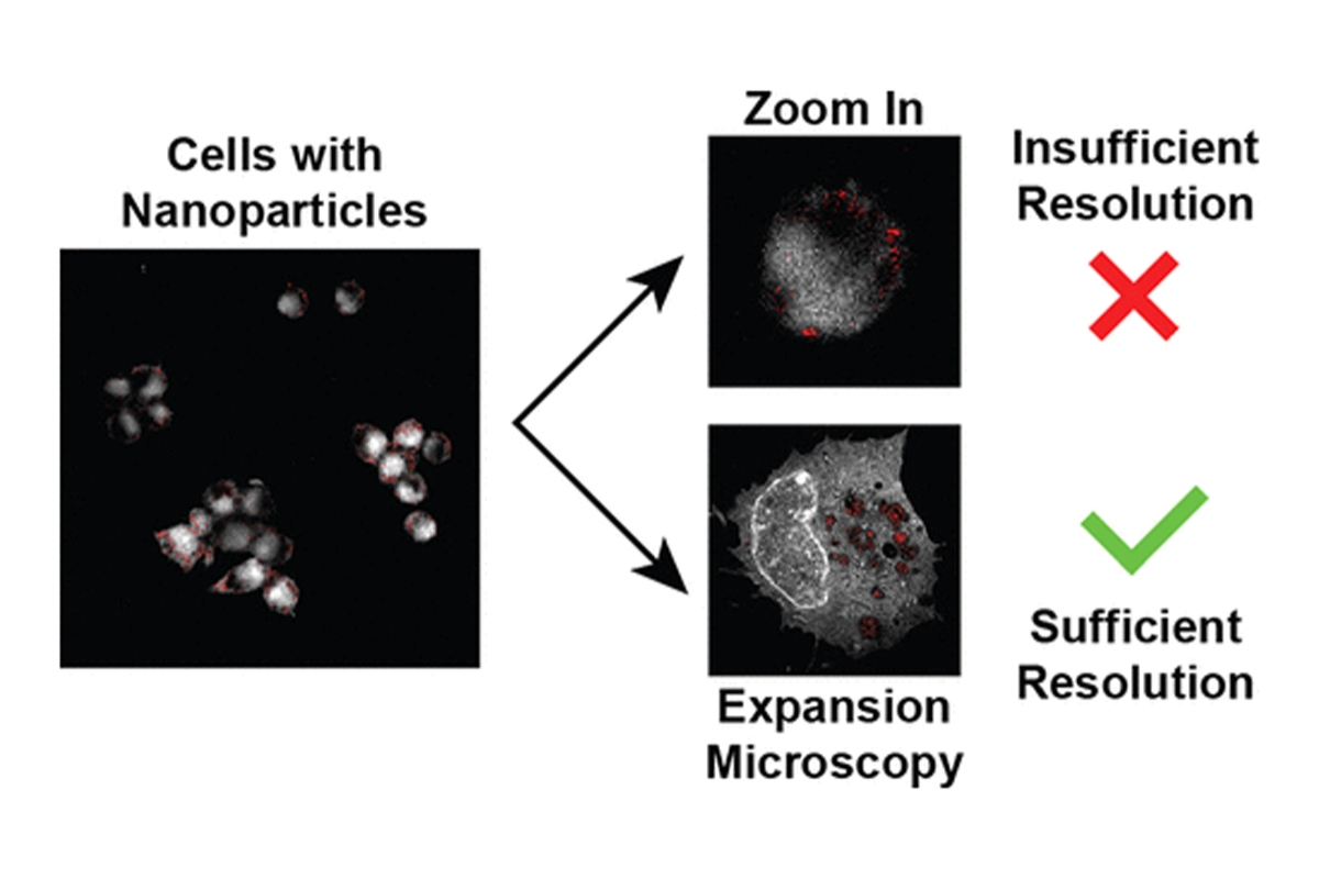
“To see nanomedicines in biological samples, researchers either use electron microscopy, which provides excellent spatial resolution but lacks 3-D imaging capabilities, or optical microscopy, which achieves excellent 3-D imaging, but exhibits relatively low spatial resolution,” Wilhelm said. “We demonstrate that we can perform 3-D imaging of biological samples with electron microscopy-like resolution. This technique, called super-resolution imaging, allows us to see nanomedicines inside individual cells. Using this new super-resolution imaging method, we can now start to track and monitor nanoparticles inside cells, which is a prerequisite for designing nanomedicines that are safer and more efficient in reaching certain areas within cells.”

super resolution nanoparticles

The researchers applied a 3-D super-resolution imaging technique known as expansion microscopy which involves embedding cells within swellable hydrogels. Like water-absorbing materials used in diapers, the hydrogel materials physically expand up to 20-fold their original size upon contact with water.

“This expansion enables the imaging of cells with a lateral resolution of approximately 10 nanometers using a conventional optical microscope,” Wilhelm said. “We combined this method with an approach to image metallic nanoparticles within cells. Our approach exploits the inherent ability of metallic nanoparticles to scatter light. We used the scattered light to image and quantify nanoparticles inside cells without the need for any additional nanoparticle labels.”

The authors suggest their super-resolution imaging platform technology could be used to improve the engineering of safer and more effective nanomedicines to advance the translation of these technologies into the clinic.

The article, “Quantifying Intracellular Nanoparticle Distributions with Three-Dimensional Super-Resolution Microscopy,” is available via ACS Publications. Wilhelm, the corresponding author, is an affiliate faculty at the OU Health Stephenson Cancer Center and a faculty fellow for the OU Office of the Vice President for Research and Partnerships. The first author is Vinit Sheth, a doctoral student in the Stephenson School of Biomedical Engineering.