The overarching objective of this Administrative Core is to (1) build a well-integrated administrative infrastructure to effectively coordinate mentoring and oversight of the promising research project leaders (RPLs) and pilot project investigators in conducting their research projects and career development, (2) implement the selection and evaluation criteria for sustaining the pool of promising RPLs, and (3) support the research resource supporting cores to enhance the medical imaging related translational cancer research activities of the RPLs and other researchers supported by this COBRE.

This Core aims to implement an interdisciplinary management plan to build an optimal infrastructure for supporting medical imaging related translational cancer research projects, including providing service using the existing imaging equipment and developing new investigative imaging testbeds and quantitative image feature analysis tools. Thus, this Core is not merely a collection of the existing imaging equipment in a distinct space; it also allows us to fully network our current research expertise to provide experienced support as we move from basic studies into more innovative translational research projects that could benefit cancer patients in the future.
This Core serves as an important bridge to link biomedical imaging engineering-based research and the translational clinical cancer research. The Core will provide research project leaders (RPLs) with the regulatory and biostatistics support to ensure full compliance with all federal regulations and achieve high scientific rigor in performing their research projects. The Core will also build a unique integrated medical imaging informatics infrastructure or a centralized database platform that contains the annotated radiological and pathological medical images along with the curated clinical outcome data and the biomarkers that link to the biospecimen metadata.
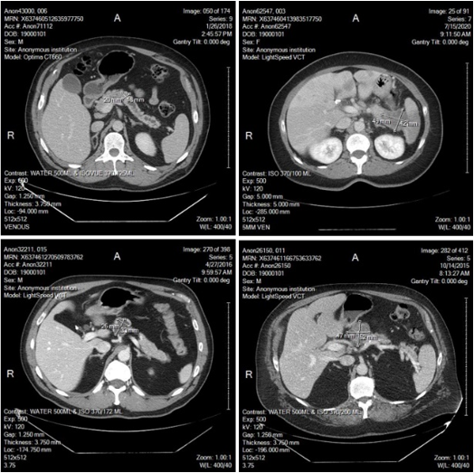
Images on the right show examples of CT images recently annotated by a radiologist to support medical imaging research to identify quantitative image markers to help diagnosis of pancreatic tumors. Based on RECIST guidelines, two maximum perpendicular diameters of the central tumor region are measured and marked (as shown in images). In these four images, two tumors (on the top) are verified pancreatic adenocarcinoma, while two tumors (on the bottom) are intraductal papillary mucinous neoplasm (left) and serous cystadenomas (right), which currently are benign, but have a risk of progressing to pancreatic cancer.