Skip Navigation

How to Study ‘Big’ Molecules Without Breaking Them Apart

Inside OU

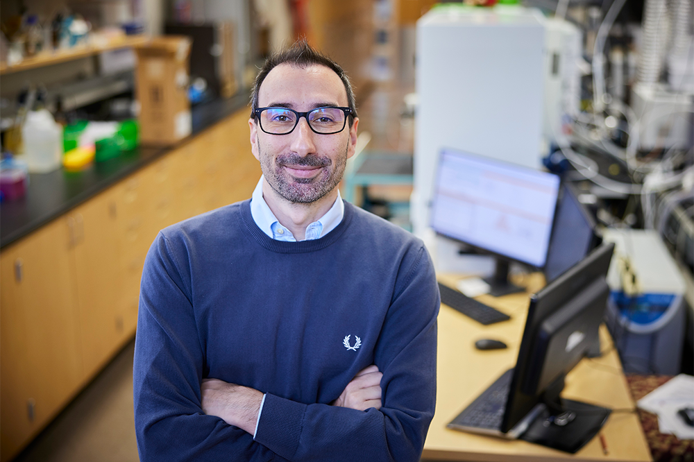
portrait of Fornelli in the lab
Luca Fornelli

How to Study ‘Big’ Molecules Without Breaking Them Apart


Just like seeing the forest through the trees, studying a whole, intact molecule has incredible scientific benefit but is difficult to do without the very latest technology.  

Luca Fornelli, an assistant professor of biology in the Dodge Family College of Arts and Sciences at the University of Oklahoma, has received a five-year Maximizing Investigators' Research Award, or MIRA, from the National Institutes of Health to increase the efficiency of National Institute of General Medical Sciences funding by providing researchers “with greater stability and flexibility, thereby enhancing scientific productivity and the chances for important breakthroughs.”

Fornelli studies proteomics, the science of studying the entire ensemble of proteins inside certain cells or tissues. Proteins are large, complex molecules that play many critical roles in the body. They do most of their work in cells and are required for the structure, function and regulation of the body's tissues and organs. However, Fornelli said, “A protein doesn't exist per se.” Instead, there are many different forms of proteins called proteoforms.

“The standard way proteomics is performed requires that a protein be deconstructed – chopped into pieces so that those smaller components can be studied to try to infer information about the original molecule,” Fornelli said.

This method, known as “bottom-up” or “shotgun” proteomics, is used because molecules are often quite large and difficult to study as a whole. 

Fornelli’s lab specializes in an approach called “top-down proteomics” which looks at the protein forms as they originally are. This is important because it helps scientists understand a molecule’s different modifications that may alter the activity of protein forms in tissues or cells. An example is the COVID-19 virus' spike protein.
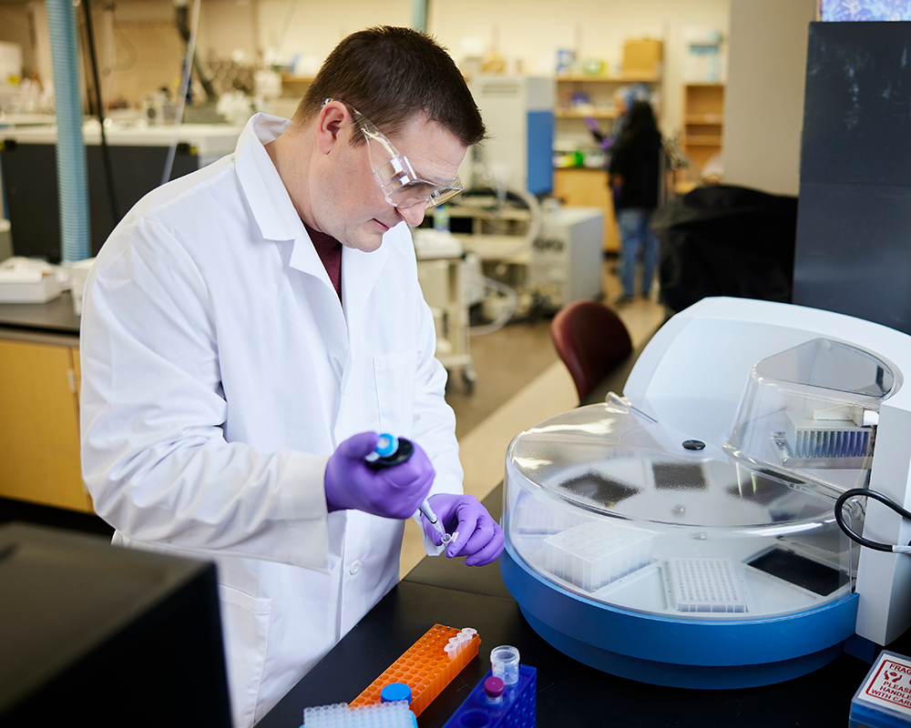
Klien, dressed in lab gear, prepares samples for testing
Postdoctoral researcher Jake Kline, Ph.D., demonstrating the KingFisher Flex equipment acquired through the OU Office of the Vice President for Research and Partnerships’ Strategic Equipment Investment Program.

The COVID-19 spike protein has multiple sites of a very specific sugar modification called glycosylation. Using the more common bottom-up method of studying proteomics, scientists would be able to tell where the sites of modifications are and potentially how they are modified.

Fornelli said, “Once you have chopped your protein into pieces, you cannot reassemble it together and make sure which piece went with which other … and there are some modifications that, once on a protein, exclude some others – or some that can be added on top of a molecule only if others are already present. So, unless you have the possibility of looking at this proteoform as an intact molecule, it's basically impossible to tell how these modifications interact.”

In other words, scientists could know where modifications occurred and identify single modifications, but they could not determine how the components combine. Whereas with the technology Fornelli is working to develop, scientists would be able to see the full picture – how the different modifications go together.

Fornelli’s submission for the MIRA award used data collected through an instrument acquired by the University of Oklahoma through its 2021 Strategic Equipment Investment Program offered by the university’s Office of the Vice President for Research and Partnerships. The $50,000 investment purchased a state-of-the-art instrument for automated, high-throughput purification of biomolecules and is used by molecular scientists in multiple fields. Fornelli said this instrument improves how researchers can compare multiple interactions simultaneously.

“When you study diseases, especially – but also with basic biology – you normally don't have just a binary A versus B type of comparison, but you need multiple controls,” Fornelli said. “In our case, we want to monitor all the changes in the proteoforms that we can detect, and we want to do that with multiple biological replicates to have statistical power to say, ‘yes, this condition is caused by the presence of this or that proteoform’ or, ‘yes, the drug is actually working; it didn't happen once, just by chance.’ So, this is a device that can allow you to get high-throughput results, and that's very important when it comes to both the basic biologists studying a certain mechanism in cells, as well as to perform translational studies.”

Now, with the support of the MIRA funding, Fornelli wants to ensure his lab can qualitatively and quantitatively analyze proteins that are intact even if they're very big.

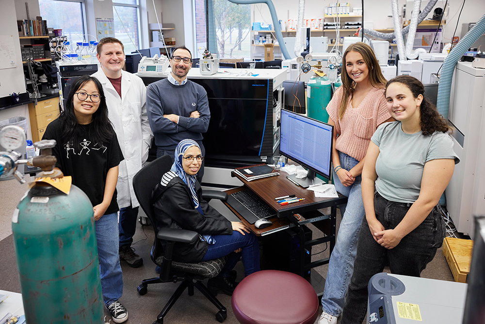
Posed photo of the group within the lab
From left: Linda Lieu, Jake Kline, Luca Fornelli, Amal Eltobshi, Alyssa Hargis, Grace Goodwin.

“The majority of our problems when we ran top-down experiments come with proteins that have a certain molecular weight,” he said. “On top of that, over the years, mass spectrometry has evolved so that now we can see complexes of different proteins. This is very important because we can study the structure of these complexes, and these complexes represent the actual form used by proteins' work in cells, as little molecular machines.”

Specifically, Fornelli and a collaborator have recently been studying how chaperone proteins work based on the set of modifications that they have. Chaperones are a group of proteins that are functionally related and can serve as catalysts for how proteins can aggregate or “fold” together when a cell experiences physiological or stress conditions.

“They interact with this or another protein, depending on how they are modified, so essentially they generate not one but a myriad of different complexes depending on how their surface is modified,” he said.

Two of the chaperone proteins they are looking at are very large in terms of top-down proteomics, making them difficult to study and even more so when they interact with other proteins.

Aside from basic biology, developing techniques for characterizing large intact proteins could prove useful for the development and testing of therapeutics.

“When you think about biologics, or new protein-based drugs, this type of technology would be very important because we wouldn't otherwise be able to look with a very good level of molecular detail at the way these molecules are produced, to know if there is a real correspondence between the intended design and the actual molecule. And then, later on, how they survive in blood,” he said. “Because, of course, when they are injected in a patient, they are in an environment where they can start being modified. So, knowing the modifications will be important due to their direct consequences for the efficacy of the therapy and health of the patient.”

The project, A multi-level mass spectrometry pipeline for the analysis of whole proteoforms and their complexes, is funded by the National Institutes of Health, award no. 1 R35GM147397-01.


This article was originally published by the Office of the Vice President for Research and Partnerships.

Article Published: Wednesday, November 30, 2022