Application Information & Scheduling Contacts
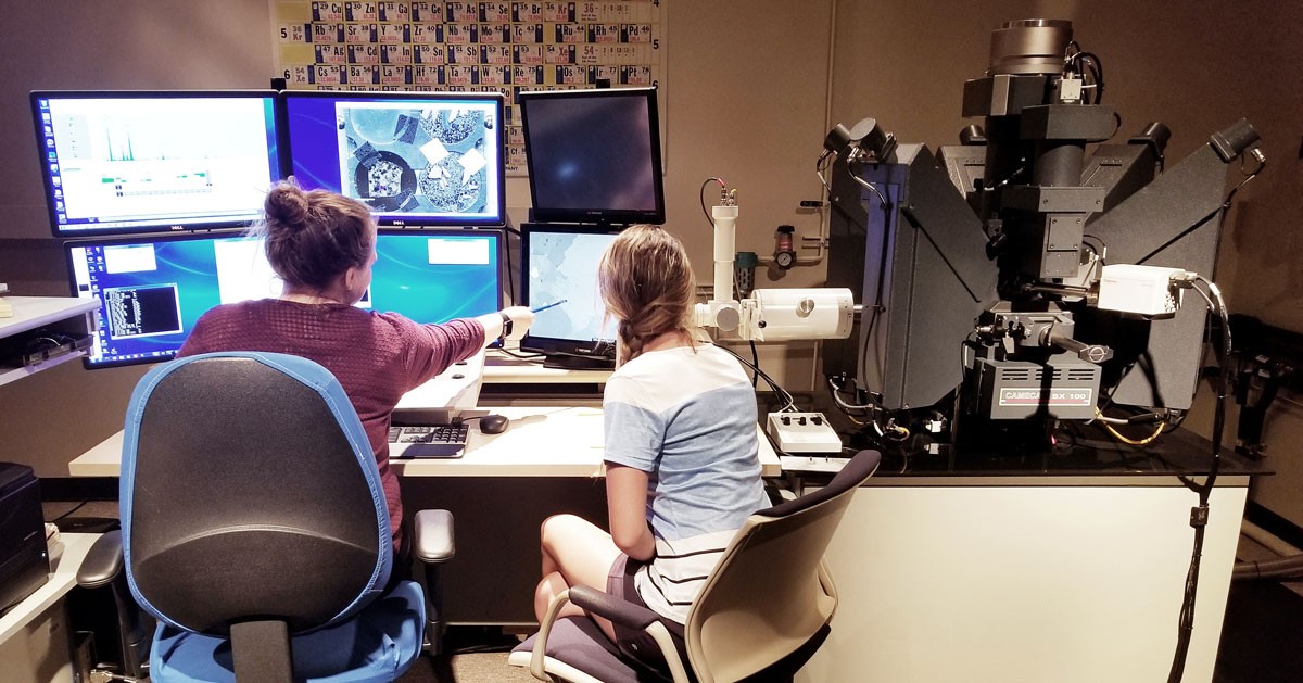
Electron Microprobe Laboratory
Sarkeys Energy Center
100 E Boyd St, Room E106
Norman, Oklahoma 73019-1009
Dr. Lindsey Elise Hunt
Laboratory Operator
Sarkeys Energy Center, Room E110
P: 405.325.2642
F: 405.325.3140
Email: lehunt@ou.edu
Related Links
Electron Microprobe Laboratory
This facility is available to clients from academia, government, industry, and the private sector for rapid chemical and spatial characterization of solid samples using electron microbeam methods. The facility is based upon a fully computer-automated CAMECA SX100 electron probe micro-analyzer that is equipped with five wavelength-dispersive x-ray spectrometers, integrated energy-dispersive x-ray analyzer, standard SEM imaging capabilities, and cathodoluminescence detector. A full-time staff is available to conduct analyses and for consultation or instruction.
Electron microbeam techniques are extremely versatile, with the capability for:
- Full quantitative analysis of elements with atomic numbers from 5 (boron) to 92 (uranium)
- Qualitative to semi-quantitative analysis of major to minor elements in seconds
- Analysis of particles or areas down to ~2-5 mm in diameter, in samples up to several cm2
- Imaging capabilities including secondary electron, backscattered electron, x-ray intensity, absorbed current, and cathodoluminescence signals that are acquired and stored in digital format
- On-line and off-line data reduction, statistical analysis, spreadsheets, graphics, and qualitative or quantitative image analysis
Applications:
Essentially any solid material that is stable under high vacuum can be analyzed and/or imaged with the microprobe. These include, but are not limited to: (1) minerals (rocks), salts, and synthetic crystals; (2) glasses and ceramics; (3) bone and shells; (4) coal and dried organic tissues; (5) metals and their corrosion products; and (6) electronic components. Thus, the electron microprobe can be applied to studies involving the composition, morphology, or abundance and distribution of phases within a wide variety of solid materials.